


To date, science has identified about 280 species of worms that can grow and live in the human body and parasitize on various organs and tissues. The frequency of human worm infections depends on the climatic and socio-economic conditions of specific areas (in underdeveloped countries, especially in tropical and subtropical areas, the level of parasitic infections is higher than in economically developed countries).
Ways of human infection with helminths
- Biohelminthiasis (infection from animals).
- Infectious helminthiasis (transmitted from person to person).
- Geohelminthiasis (diseases caused by parasites that carry out one of the life cycles on Earth).
Factors affecting the manifestations of helminthiasis
- the way a parasite enters the body;
- degree of adaptation of helminths to the human body;
- Population density (number) of parasitic individuals;
- Worm habitat (tissue parasites live in the thickness of soft tissues, and luminal ones live in the lumens of hollow organs). Some helminths in different phases have both luminal and tissue forms. The larval and developmental stages of worms, as a rule, lead to more pronounced pathological changes.
In the absence of re-infection, the number of adult parasites in the human body does not increase. This feature significantly distinguishes helminthic invasions from diseases caused by bacteria, viruses, fungi and protozoa.
Worms in humans: symptoms
Helminthiasis is a disease characterized by two stages of the course (acute, two weeks to two months) and chronic (several months to several years).
Symptoms of the acute phase of helminthiasis
The first symptoms of the disease may appear at different times (after 2-3 weeks at most, with ascariasis - after 2-3 days, and the incubation period with filariasis can last 6-18 months).
The most characteristic symptom in the acute phase of parasitic heat is an allergic reaction (antibodies are produced to the antigens of migrating parasitic larvae). People infected with worms often develop itchy rashes on the skin, prone to recurrent course, enlarged regional lymph nodes, and may develop generalized or local edema, muscle and joint pain. Migrating parasitic larvae can also cause chest pain, coughing, choking attacks, stool disorders, nausea and vomiting.
At the same time, the acute stage of helminthiasis may be accompanied by more serious disorders (severe forms of pneumonia, hepatitis, allergic myocarditis, hepatosplenomegaly (enlarged liver and spleen), meningoencephalitis).The number of eosinophils in the blood increases (eosinophilia) and the normal quantitative ratio between protein fractions is disturbed (dysproteinemia).
Symptoms of chronic helminthiasis
The symptomatology of the chronic phase directly depends on which organ is "inhabited" by parasites, as well as their size and number play an important role.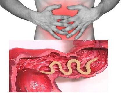 Thus, the disease may be asymptomatic when parasitized in the intestines of individuals (except in cases of infection with very large parasites). Characteristic symptoms of the chronic stage of intestinal helminthiasis are dyspeptic diseases. Asthenoneurotic and pain syndromes are more common in children. Mass invasion of roundworms can lead to the development of intestinal obstruction, obstructive jaundice and pancreatitis.
Consumption of all the substances necessary for vital functions from the body of the host, helminths cause digestive disorders, the absorption of vitamins, minerals, carbohydrates, proteins and fats. At the same time, worm waste interferes with the normal intestinal microflora and reduces the body's immune system.
People suffering from helminthiasis have a significantly increased risk of malignant tumors due to weak immunity and the process of cell division (as a result of the constant regeneration of tissues damaged by parasites).
Types of helminths parasitizing the human body
The causative agents of human helminthiasis are 2 types of worms: roundworms (nematodes) and flatworms (tapeworms and liverworms).
Round Worms
Pinworm
The parasites that cause enterobiosis are small (up to 10 mm) thin hollow worms of grayish-white color. The infection is edible (orally). This is due to dirty hands. The parasite's eggs are found in soil, in the wool of infected animals, in unwashed vegetables and fruits, and so on. Can be. At the same time, spontaneous infections with enterobiasis often occur (especially in children), itchy areas and subsequent ingestion of eggs.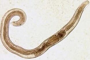 Pinworm larvae develop in the digestive system within two weeks. When it matures into an adult, the worm parasitizes the lower and upper parts of the colon.
In the larval stage, pinworm also begins to damage the host's body, producing enzymes that irritate the intestinal wall and cause an inflammatory process to develop. Adult parasites attach or penetrate deeper layers of the intestinal mucosa, disrupting its integrity and helping to close secondary bacterial infection. In the case of perforation of pinworms in the wall of the small intestine, peritonitis may develop. Also, due to irritation of intestinal receptors, motor and secretory functions of the gastrointestinal tract are impaired, gastroduodenitis, enteritis, etc. Prolonged enterobiasis in childhood can lead to neurological disorders and retardation in physical development.
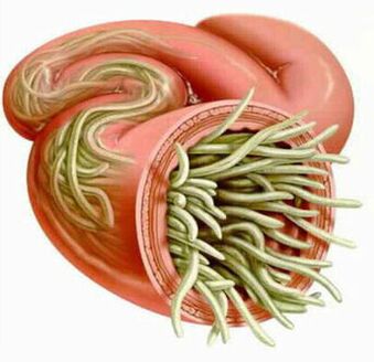
Ascaris
Ascaris is a reddish-yellow parasite in the form of a large spindle, reaching 40 cm (female) and 15-25 cm (male). Without suction cups or other fastening devices, the roundworm can move independently towards food masses. Eggs laid by the parasite's teeth are excreted in the feces.
Ascaridosis occurs when adult eggs are washed with water or swallowed with unwashed vegetables and fruits that contain soil particles. Once the eggs enter the intestines, the adult larvae emerge. They then penetrate the intestinal wall, enter the heart through the bloodstream, and from there enter the lungs. Through the alveoli of the lungs, the roundworm larvae re-enter the oral cavity from the respiratory tract. After repeated washing, the parasite reaches the small intestine, where it reaches adulthood. The worm lives for 12 months, then dies and is excreted in the feces. Both one and hundreds of individuals can live in the intestines of an owner.
Roundworms, which have the ability to move in a spiral in the intestinal phase of their bodies, can penetrate even the narrowest holes. This feature of the parasite often leads to the development of quite serious complications (obstructive jaundice or pancreatitis). Allergens secreted by roundworms can cause severe allergic reactions. Many adults can cause intestinal obstruction, and worms that enter the respiratory tract can sometimes cause suffocation.
Vlasoglav
Vlasoglav, the causative agent of trichocephalus, is a white helminth that parasitizes in the early part of the large intestine and reaches a size of 4-5 cm, the parasite feeds on the blood and tissues of the rectal mucosa.
Corn eggs laid by the female on the intestinal wall are excreted in the feces. Their development takes place in the environment (preferably in the soil). Eggs grown in the larvae of the parasite enter the body through food, dirty hands, water or unwashed vegetables and fruits.
Trichocephalus is asymptomatic with a small number of worms. In a severe stage (mass invasion), the patient develops abdominal pain, sometimes severe diarrhea accompanied by rectal prolapse. This condition is most common in debilitated children. With a moderate stage of trichocephalus, a child's growth retardation is possible.
Trichinella
The causative agent of trichinosis is a small round helminth 2-5 mm long. The infection occurs when eating poorly roasted meat (pork, bear meat, boar).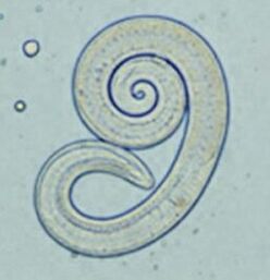 The larvae of the parasite, which penetrate the intestines, mature in 3-4 days to the level of a sexually mature individual. The life of the worm is 40 days, after which the parasite dies. By piercing the intestinal wall, the larvae enter the bloodstream and are located in the muscles and carried to all organs of the human body. In this condition, the respiratory and facial muscles, as well as the flexor muscles of the limbs, are most affected.
In the first days after the occupation, patients complain of abdominal pain. Then, after about 2 weeks, the body temperature rises to 39-40 C, itchy rashes appear on the skin, muscle pain develops and the face swells. During this period, in the case of a mass infection, the risk of death is high. After about a month, the patient recovers. The parasite is encapsulated in a spiral, and then dies within two years.
Hookworm and nekator
These two parasites have similar biological characteristics and the diseases they cause. In this regard, it is customary to combine them under a common name (hookworm). Worms up to 10-15 mm in length are parasitized at 12 p. m. intestine. It should be noted that this is one of the most common, but also very rare parasites. Worm larvae enter the human body through the skin when they come in contact with contaminated soil. In addition, they enter the bloodstream and, like roundworms, migrate to the lungs, where they then enter the digestive system through the bronchi, along with mucus. Hookworm parasitizes in the intestine, attaches itself to the intestinal wall. The blood-only parasite bites the blood vessels that penetrate the mucous membrane and injects the anticoagulant component. On average, an adult can receive 0, 05-0, 35 ml of blood per day. Therefore, the most characteristic symptom of this helminthiasis is iron deficiency anemia, as well as changes in the ratio of protein fractions (dysproteinemia).
Flatworms
Wide Ribbon
This is one of the largest helminths, reaching a length of 10-20 meters. The disease caused by this parasite is called diphyllobotriasis. The developmental period of the worm begins with freshwater fish or crustaceans. The larva enters the human body with the egg or infected fish fillet, which is the final owner of the tapeworm. The parasite attaches to the wall of the small intestine and grows in an adult for 20-25 days.
Difillobotriasis occurs against the background of anemia of the digestive system and B12 deficiency.
Liver fluke
The parasite that causes opisthorchiasis is a flat worm 7-20 mm long. It should be noted that more than 50% of cases of hepatitis (also called feline hepatitis) occur in Russians. The larvae of the parasite begin to develop after the eggs enter the fresh water (from the snails that swallowed them). Then they penetrate the body of the fish (carp, crucian carp, grass, roach). Human infection occurs when eating contaminated fish meat that has not received adequate heat treatment. The larvae of the liver fluke from the small intestine penetrate the bile ducts and gallbladder, where they are secured with two suction cups.
In the acute stage of helminthiasis, the patient has pain in the upper abdomen, fever, nausea, muscle aches, diarrhea and skin rashes. Chronic opisthorchiasis manifests itself with symptoms of hepatitis, inflammation of the bile ducts, cholecystitis, digestive disorders, nervous disorders, weakness and increased fatigue. The parasite causes irreversible changes, and the patient does not suffer from chronic inflammatory processes and functional disorders even after expulsion.
Beef and pork tapeworm
The length of these parasites, which are almost identical in structure, reaches 5-6 meters. Infection with teniarinhoses and teniasis occurs due to the consumption of meat from cattle or pork (one of the intermediate forms of helminthiasis) infected by Finns. Presented in the form of whitish bubbles up to 0. 5 cm in size, living Finns attach to the wall of the human small intestine and become an adult in 3 months. The tape parasite, which consists of more than 2, 000 segments, is constantly growing. In this case, the last segments of the egg break down and move independently along the large intestine to the anus, then crawl out of the anus or are excreted with the feces. The most characteristic symptoms of helminthiasis are disorders of the digestive system.
Echinococcus
For this parasite, man is an intermediate host. The worm parasitizes the human body in the form of Finns. The final owner of the echinococcus is a wolf, dog or cat.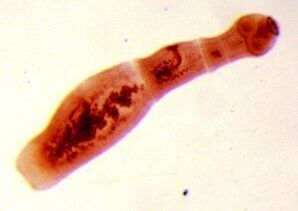 The infection comes in contact with animals and environmental objects planted with Echinococcus eggs. Once inside the gut, oncospheres (six-hooked larvae) develop from them. They enter the bloodstream from the intestines and travel throughout the body.
The worm's "favorite" parasitic sites are the liver and lungs. By settling in these organs, the larva develops into a fin (echinococcal cyst), which gradually increases in size and begins to destroy nearby tissues. Echinococcosis is often mistaken for a tumor of benign or malignant origin. In addition to mechanical effects (compression of organs and blood vessels), sometimes rupture of echinococcal cysts occurs. This condition can lead to toxic shock or the formation of many new cysts.
Alveococcus
This parasite, which is considered a type of echinococcus, is the cause of one of the most dangerous helminthiases (alveococcosis), similar to the severity of cirrhosis and liver cancer. Infection occurs when oncospheres (eggs with adult larvae) enter the intestine. There, the embryo leaves the egg and enters the bloodstream by penetrating the intestinal wall. In addition, the parasite spreads through the bloodstream to all tissues and organs of the body (most often localized in the liver). The main stage of development begins with the larvae (a multi-chambered bubble, laurocyst is formed). Each cell has an embryonic head of the parasite, which continues to develop gradually. Laurocysts are very aggressive formations that grow continuously due to expanding bubbles and also have the ability to grow in the liver as cancerous metastases. Necrotic changes due to disorders of blood vessels are accompanied by necrotic changes in nearby tissues. Alveococcus, which spreads to nearby structures, forms fibrous nodules with the insertion of multi-chambered bubbles. This condition can last for several years and therefore requires mandatory surgery.
Diagnosis of helminthiasis
Diagnosis of helminthic invasions includes the following activities:
- Get a comprehensive history to help find possible causes of infection;
- feces, blood, intestinal contents 12p, rectal and perianal mucus, muscle tissue, pulmonary sputum, bile laboratory tests. Eggs, segments or parasitic fragments may be detected during the analysis. At the same time, the increased content of eosinophils in the blood is a signal of the presence of helminthiasis.
- Serological tests are performed when diagnosing diseases caused by larval stages or tissue parasites (ELISA, RSK, indirect agglutination test, immunofluorescence analysis, etc. ).
- Ultrasound, CT and endoscopic examinations are prescribed to detect helminths affecting liver tissue.
Human worms: treatment
In the acute stage of parasitic infection, the patient is prescribed detoxification and desensitization therapy. In severe cases of the disease (liver trematodes, trichinosis), glucocorticoids are used according to medical indications.
Special anthelmintic chemotherapeutic agents are prescribed as drugs of special therapy, taking into account the nature of the pathogen.
In parallel, the patient is recommended to take antihistamines and enterosorbents. The final stage of treatment involves the use of probiotics that normalize the intestinal microflora.
A special frugal diet is also prescribed (food should be digested and low in fat).
During anthelmintic therapy, the patient is required to adhere to strict personal hygiene (to prevent re-infection). At the same time, for many helminthiases, all family members and those in constant contact with an infected person should be treated.
Prevention of helminthiasis
- Protection of personal and public hygiene;
- Strict adherence to cooking technology;
- Regular examination and preventive treatment of pets;
- Thorough washing of fresh vegetables, fruits and herbs;
- Proper management of tea fish;
- Avoid raw, lightly salted and dried fish.